TL;DR
Distinct synaptic connections from the cortex to the pulvinar create distinct oscillatory patterns, with significant implications for visual processing in the cortex.
Context
The pulvinar is a thalamic nucleus located along the medial edge of the lateral geniculate nucleus. Throughout mammalian evolution, its size and differentiation have notably increased, correlating with the neocortex’s development [1]. It is by far the largest nucleus of the primates’ thalamus, and has a unique extensive (and recroprocical) synaptic connections with all visual cortical areas.
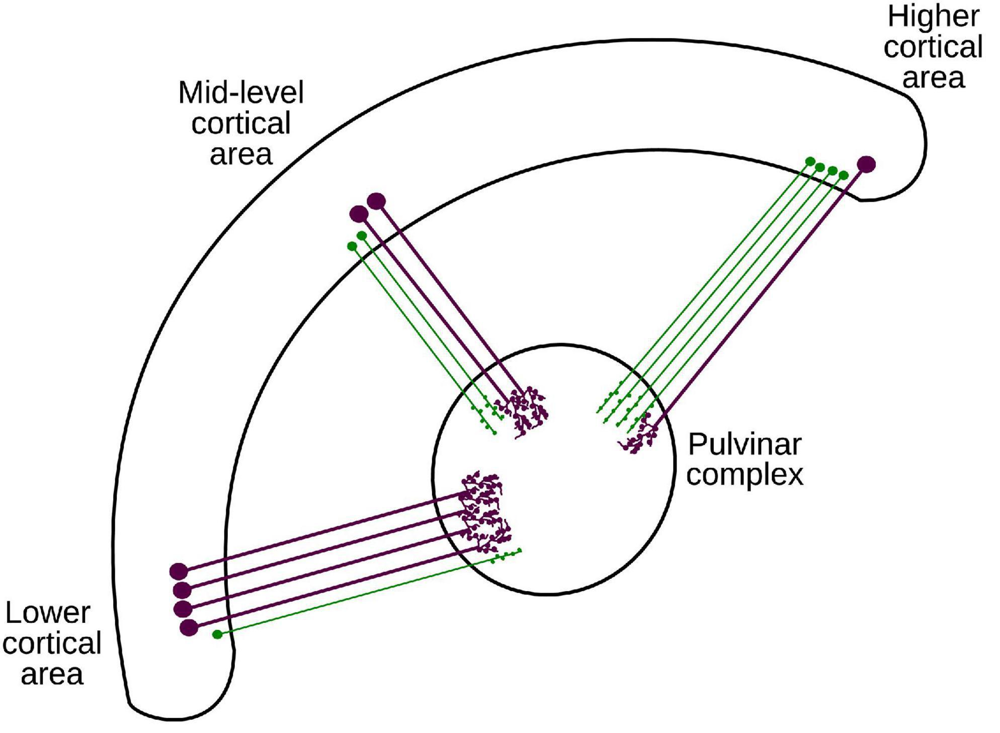
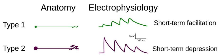
Synaptic connections to the pulvinar, however, are not uniform. Depending on the hierarchical level of the cortical area they originate from, the connections differ [2]. For instance, low-level cortical areas (e.g., primary visual cortex - V1) project synapses that cause depression, whereas higher-level areas (e.g., V4) have synapses leading to post-synaptic facilitation.
Takeaways
In this paper, we created a spiking neural network model of the pulvinar, and studied the why of varying those two types of synapses can change the activity in the pulvinar.
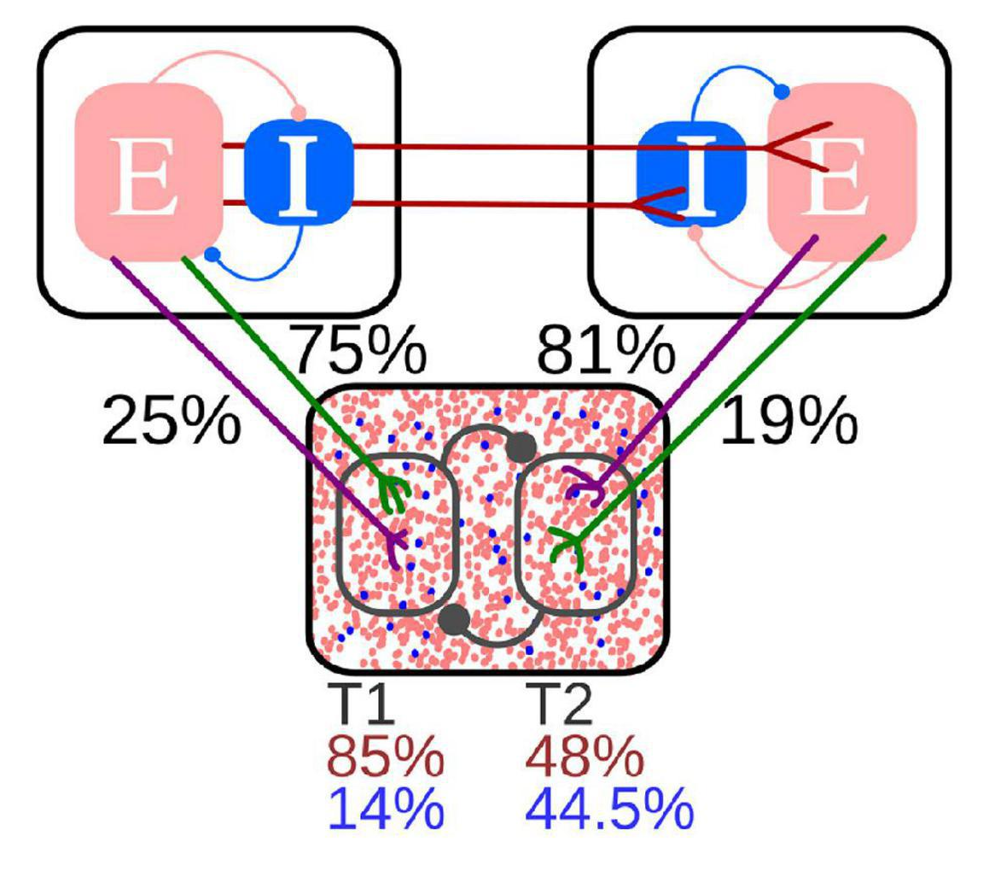
Our model demonstrated that V4 and V1 projections have opposing effects on the pulvinar’s state. When pulvinar neurons are in an alpha oscillatory state due to input from V1, V4 input shifts the response to a non-oscillatory spiking activity, and vice versa.
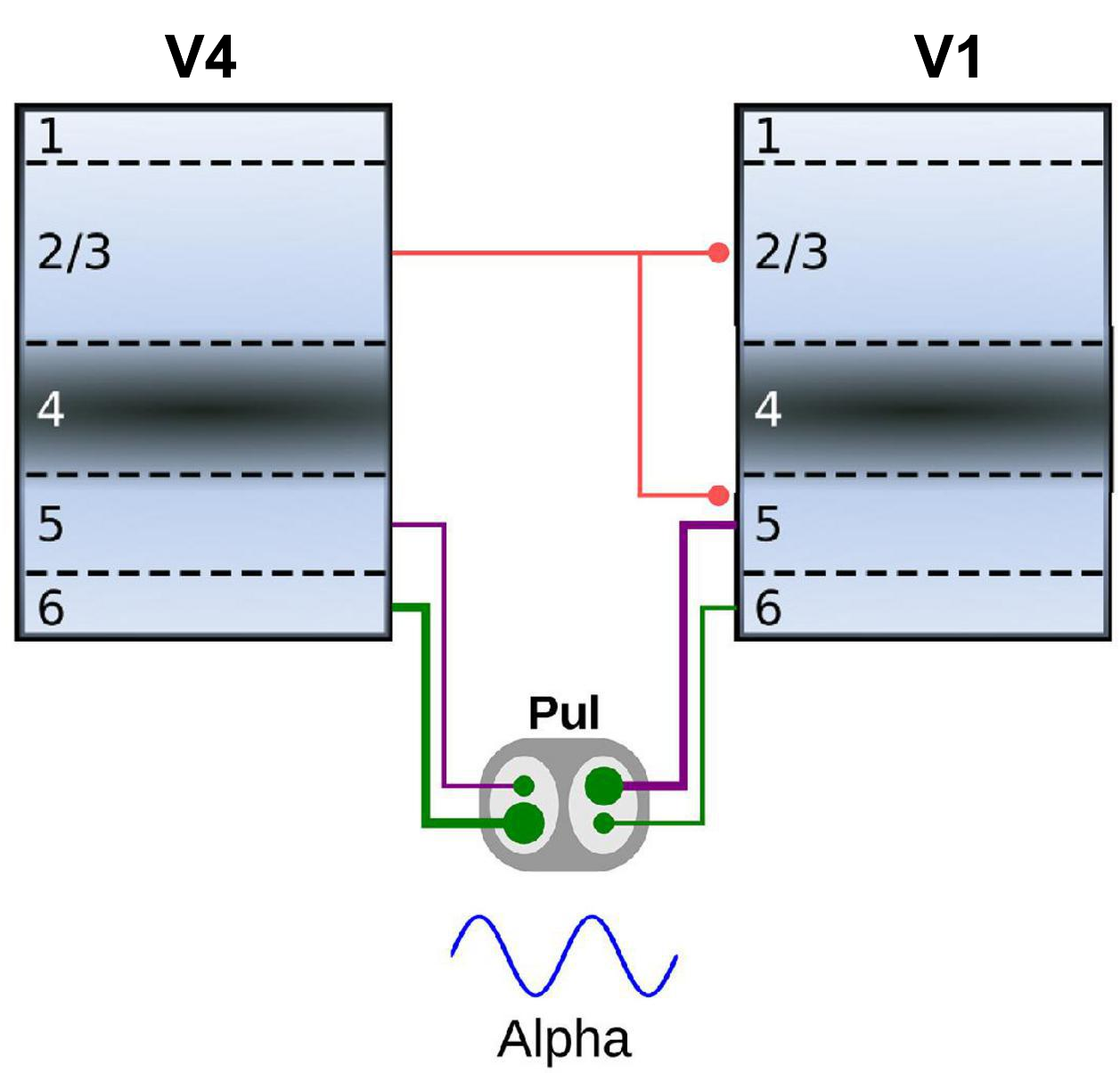
If direct connectivity between the two cortical areas is added to the model, the alpha oscillatory state generated by V1 in the pulvinar also propagates to V4. When synaptic activity from V4 reaches the pulvinar, the system transitions into the previously mentioned non-oscillatory state, closing the oscillation loop.
Relevance
The canonical view of visual processing in the cortex is that of a hierarchical communication system. In this paper, we showed that alpha oscillations, typically associated with high-to-low level (feedback) communication, can actually travel in the opposite direction by jumping through the pulvinar.
References
[1] Hutchins, B., & Updyke, B. V. (1989). Retinotopic organization within the lateral posterior complex of the cat. Journal of Comparative Neurology, 285(3), 350-398.
[2] Abbas Farishta, R., Boire, D., & Casanova, C. (2020). Hierarchical organization of corticothalamic projections to the pulvinar. Cerebral Cortex Communications, 1(1)
On a personal note
This was my very first peer-reviewed paper !
